Table of Contents
In today’s lesson we discuss women’s bone health in some detail especially as it relates to osteoporosis. We will also cover bone architecture as it relates to bone health.
Recent technological developments allow researchers to look deeper into bone architecture and determine the effects of different treatment options for osteoporosis. Researchers can examine the relationship between bone density and bone quality. This is an important development for women’s bone health.
For many years bone density has been the key metric to evaluating women’s bone health. But now we are learning more about bone quality. This is one of the more exciting areas in women’s bone health today.
You are welcome to dive deeper into the topic by going to Today’s Reading Material that I have prepared for you at the bottom of this page!
Continuing Education Course for Bone Health
I encourage you to check out the Building Better Bones online course for Physical Therapists, Physiotherapists, Kinesiologists, Athletic Trainers, Occupational Therapists and Physical Therapy Assistants.
Over 12,000 health professionals have completed this course.
“This course should be a mandatory part of all Physiotherapy training programs.” – Karen Ginsburg, Physiotherapist, Community Rehab Therapy, Hamilton, Ontario.
“MelioGuide™ is an evidence-based approach to teaching your clients postural, strength, cardio, flexibility, and balance exercises that are tailored to their level of fitness and risk of fracture.” — Holly Bonasera, MPT, GCFP, Licensed Physical Therapist, Certified Feldenkrais Practitioner, Blair, Nebraska
“The MelioGuide Building Better Bones course was exceptional — both for me as someone with Osteoporosis and as a Health Care Professional who treats clients with Osteoporosis.” — Alison Krausen, Occupational Therapist, Genesis Rehab Services, Pennsylvania
Understanding Women's Bone Health
Women’s bone health is a major health issue. Half of all women over the age of 50 are going to have a fracture in their remaining lifetime.
Today we’re going to talk about bone density and bone quality, and how they affect someone’s fracture risk. In the people that fracture, studies have shown is that half of those fractures occur with people that have relatively good bone mineral density scores.
Bone Quality and Bone Health
Why do some people fracture while others do not? Most recent studies have shown that it is related to bone quality. Recent diagnostic tools have allowed us to look more deeply into the quality of bone, and that tool is called a peripheral quantitative computer tomography (PCQT).
Peripheral Quantitative Computer Tomography
Unfortunately for many people, peripheral quantitative computer tomography is expensive and not readily available. There are few peripheral quantitative computer tomography machines accessible to the general public. As a result, most of your clients will never have the opportunity to have a PQCT done.
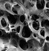
However, even without access to this measurement, I think it’s very important that you know about bone quality. The quality of bone is defined as a combination of the microarchitecture of the bone and a microstructure of the bone.
The microarchitecture of the bone is a combination the type of rods and cones, the shape of the trabeculae, and the thickness of the trabeculae. The microstructure reflects the quality of the collagen as well as other bone mineral matrix.
At the 2010 American Society of Bone Mineral Research conference, a number of researchers presented studies on the use of the peripheral quantitative computer tomography tool to look at whether or not women who fractured had different bone makeup than women who didn’t fracture but with the same bone mineral density and in the same age group.
Bone Quality and Bone Density
In the women who fractured, even though they had the same bone mineral density and were of the same age group as the women who did not fracture, it was shown by the peripheral quantitative computer tomography that there were fewer plates and fewer connections between the plates and rods. This means that there were less trabeculae and less bone in the women who fractured. A bone mineral density test does not provide this information.
A bone mineral density test strictly just looks at the density of the bone all the way through. A BMD or DEXA test is unable to discriminate between trabecular bone or cortical bone, or between thickness of the trabeculae themselves.
In the last decade the World Health Organization recognized that their old definitions of osteoporosis and osteopenia were not sufficient to identify who should be on medication based on their fracture risk.
FRAX
As a result, they developed FRAX — a questionnaire that looks at a lot of other elements that determines women’s bone health. It examines variables beyond their age, family history, calcium intake, vitamin D intake, and a number of other things such as smoking. I have an article that explains the FRAX.
Bone Architecture
We now have a better understanding of women’s bone health, bone architecture and its relevance to bone quality.
Scientists can now look at the bone architecture through the use of High Resolution peripheral Quantitative Computed Tomography (HR-pQCT). This allows researchers to study the quality of bone structure at the micro level and, hopefully, understand what activities promote improved bone quality and are effective strategies for osteoporosis prevention and treatment.
Two recent studies provide more insight into trabecular architecture as well as the mechanical behaviour and fragility of bone. In the following sections, I provide a summary of each of these two studies.
Trabecular Microarchitecture and Mechanical Behaviour of L3
At the American Society of Bone Mineral Research (ASBMR) meeting in Toronto in October of 2010, research outlining the contribution of trabecular microarchitecture to the mechanical behaviour of L3 was presented. (1)
Before I get into the details of the study and its results, I need to provide some definitions for the reader.
Trabecular Plats and Trabecular Rods — Identification Based on Shape
Trabecula fall into two broad categories based on the shape of the trabecula.
These are defined as:
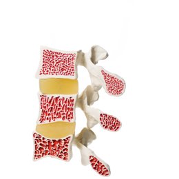
- Trabecular Plates: Trabecular Plates are the trabecula that are thicker and more “plate-like” in shape. In the image above, the plates are the wide, flat light colored parts of the bone. Since they are wider and thicker than the rod-like connections, they provide more support and contribute to the overall quality of the bone structure.
- Trabecular Rods: Rods are the trabecula that are “rod-like” in shape. With loss of bone mineral density, the trabecula that are plate-like can become rods as they become thinner. This change can reduce the quality of the trabelula microarchitecture. In the image to the right, the rods are the light colored, thinner rod-like connections.
Bone Architecture — Orientation
Both rods and plates occur in different orientations. The following are the three orientations:
- Transverse — Horizontal alignment
- Oblique — Oblique alignment
- Longitudinal — Vertical alignment
Bone Architecture — Segmentation
Morphological analysis of individual trabeculae segmentation can be broken into the following categories:
- Plate/rod volume fraction
- Axial bone volume fraction
- Plate/rod number density
- Plate/rod thickness
- Plate surface area/rod length
- Plate-plate/plate-rod junction density
Analysis
An analysis of L3 was undertaken from 21 fresh donors. The average age was 76 years and the group was composed of 10 men and 11 women. Bone mineral content (BMC, g) and bone mineral density, (BMD, g/cm2) were assessed by lateral DXA.
Two vertically oriented virtual biopsies were obtained – one from the anterior vertebral body and one from the posterior vertebral body. Mechanical behaviour was assessed using static compression testing involving the following three metrics:
- Failure load (N)
- Stiffness (N/mm)
- Work to Failure (N.mm)
Results & Conclusion
Trabecular architecture was found to have greater deterioration in the anterior versus posterior region. Researchers found that BMD explained up to 44% of the variability in vertebral mechanical behaviour. The microarchitecture explained up to 66% of the variability in vertebral mechanical behaviour. The combination of bone mass (BMD) with microarchitecture explained up to 86% of the variability in vertebral failure load.
These findings explain why flexion exercises, ADLs and other activities that increase the load on the anterior vertebral body can increase the risk and rate of vertebral compression fractures.
Microstructural Changes of Trabecular Bone
In another study (2) presented at the same American Society of Bone Mineral Research (ASBMR) meeting in Toronto, researchers explored the micro-structural and mechanical changes of trabecular bone in postmenopausal women who had fragility fractures.
Researchers compared 101 control subjects who had not fractured to 68 women who had sustained fractures from a fall or slip from a standing height or lower position. The research team discovered that the DXA scores of both groups were similar. However, the quality of the bone in the fracture group was much lower than that of the group that did not fracture.
The trabecular microstructure and the whole bone stiffness of the women who fractured was lower that that of the control group. The following characteristics of trabecular bone were found among the group that fractured:
- Reduced plate-like bone volume.
- Less plate-like structure.
- Less axially aligned trabecula.
- Longer trabecular rods indicative of more separated trabecula.
- Decreased connectivity between plates and plates and between plates and rods.
Although the BMD of the group who fractured was similar to that of the non-fracture group, the bone quality was the determining factor as to the strength of the bones.
References
- Wegrzyn J et al. Contribution of Trabecular Microarchitecture and its Heterogeneity to the Mechanical Behavior of Human L3 Vertebra. Univ. Lyon, France and Harvard, Boston. Poster presentation at the 2010 ASBMR Toronto.
- Liu XS et al. Fewer Trabecular Plates and Decreased Connectivity Between Plates and Rods is associated with Reduced Bone Stiffness in Postmenopausal Women with Fragility Fractures. Columbia Univ. NY. Poster presentation at the 2010 ASBMR Toronto.
Physical Therapy Continuing Education
To learn more about how to Physical Therapy Continuing Education, visit my page dedicated to Physical Therapy Continuing Education.
Comments
December 19, 2015 at 2:42pm
mustafa
Hi Margeret
First thanks for this precious lectures. I graduaded university last term. Now i m at beginning on my way i hope i wil reach your level a day. I willing so much develop myself in this process you and other physiothrepists like you be inspired to me.
February 19, 2018 at 8:29am
Andrea Karrs
I am really glad I signed up for this informative course. Can't wait to learn more.
February 19, 2018 at 8:30am
Andrea Karrs
I am really glad I signed up for this course. I look forward to learning more useful info in for informing and treating my folks with osteoporosis.
July 13, 2018 at 1:05am
Ana Colina
Very interested information; feel disappointed not having the tools to assess the bone quality to every one that needs it and we, the Therapists not able to help our Patients more effectively if we could count with that information , as we do with X-rays, nowadays .
THANK YOU MARGARET!, cannot wait for the next class...
September 20, 2021 at 6:29am
Aldona
I am very interested in compression fractures a profesional and as a patien because I had two compression fractures
Cuold we do something with increasing trabeculae tissue
September 21, 2021 at 7:40am
Richard Martin replies
I encourage you to read Margaret's article on compression fractures. http://melioguide.com/osteoporosis-treatment/treat-compression-fracture/