REMS (Radiofrequency Echographic Multi-Spectrometry) is a radiation-free ultrasound-based technology, marketed under the brand name Echolight, that measures bone mineral density (BMD) and estimates fracture risk. While it offers genuine advantages, no radiation, portability, and a proprietary fragility score, a 2026 study published in Osteoporosis International found that over 90% of REMS-BMD output variance was explained by the patient’s age and weight alone.
DEXA remains the clinical gold standard for osteoporosis diagnosis and treatment decisions, and the majority of physicians will not prescribe treatment based on REMS results.
Below, I’ll walk you through what REMS is, how it compares to DEXA, what the latest research shows, and what you should do if you’ve already had a REMS scan.
My Position on REMS Bone Scan
Many of my readers and clients have asked me about REMS, whether they should get a scan, whether it’s better than a DEXA, and whether I recommend it. In this post, I try to give you a balanced and impartial analysis of the technology based on the latest research and my own clinical experience.
- While I see valid applications for REMS, particularly in situations where a DEXA is unavailable or unreliable, the reality is that most of you already have access to a DEXA.
- The DEXA remains the gold standard used by the vast majority of clinicians, it provides image detail showing the actual state of your bones, and it is the most reliable way to monitor your bone health over time.
- A REMS reading does not provide insights beyond what a DEXA offers, and having two sets of results from two different technologies can create more confusion than clarity.
- As a clinician, I do not use REMS in my practice. The DEXA allows me to see the status of your bone in image form, identify compression fractures that might otherwise go undetected, and track meaningful changes from one scan to the next. That is the foundation I need to build a treatment plan I can stand behind.
What Is a REMS Bone Scan?
REMS stands for Radiofrequency Echographic Multi-Spectrometry. It is an ultrasound-based bone assessment technology developed by Echolight S.p.A., an Italian medical device company, and marketed in North America by Echolight Medical. The device received FDA 510(k) clearance in the United States.
You may also see this test referred to as a REM scan, a REM bone scan, or a REM bone density scan. These are common misspellings of REMS, which stands for Radiofrequency Echographic Multi-Spectrometry. There is no separate technology called a REM scan. If you have been told you are getting a REM scan, a REMS scan, or an Echolight scan, these all refer to the same ultrasound-based bone assessment described here.
The REMS Scan Procedure
During a REMS scan, a technician places an ultrasound probe on your abdomen (to scan the lumbar spine) and over each hip (to scan the femoral neck), similar sites to a standard DEXA scan. The device analyzes the backscattered ultrasound signals and compares them against a proprietary reference database. The process takes just a few minutes per site, requires no special preparation beyond fasting for several hours before the scan, and involves no radiation whatsoever.
Your REMS report will typically include:
- Bone mineral density (BMD) measured in g/cm², just like DEXA
- T-score and Z-score — comparing your BMD to a young adult reference population (T-score) and to your age-matched peers (Z-score), respectively
- Fragility Score (FS) — a proprietary number from 0 to 100, with higher values indicating greater estimated fracture risk. Echolight markets this as a measure of bone “quality” beyond density alone
What Your REMS Report Does Not Provide
What REMS does not provide is any image of your spine or hip. This is an important distinction from DEXA, which can generate a visual of your vertebral bodies.
I had a client who had a REMS scan and was excited that her bone scores were better on REMS than with her DEXA. The problem was that the REMS could not provide an image of her lumbar spine showing her compression fractures. Spine scores always appear better when compression fractures are present, because the compressed bone is denser. Without an image, this was invisible on her REMS report.
REMS Algorithm
Behind the scenes, REMS uses a two-step algorithmic process.
- First, it compares the ultrasound spectra from your scan against a set of reference models that have been selected based on your demographics, including age, sex, ethnicity, and body mass index (BMI).
- Second, it converts the resulting “osteoporosis score” into a BMD value using a separate equation that is also dependent on your age and weight. This two-layer demographic stratification is an intentional design feature, but as we’ll see below, recent research has raised significant questions about how much the ultrasound signal itself actually contributes to the final number.
REMS vs. DEXA: How Do They Compare?
If you’re researching bone density testing options, the comparison between REMS and DEXA is probably your most pressing question. Here’s an honest side-by-side look.
One important point: DEXA comes with its own well-documented limitations. Degenerative changes in the spine, bone spurs, and even aortic calcification can falsely elevate lumbar spine DEXA scores. Operator positioning matters. But DEXA’s physics are transparent and well understood, the BMD value it produces does not change if you enter a different age or weight into the system. As we’ll see next, the same cannot be said for REMS.
The majority of endocrinologists and general practitioners will not treat your osteoporosis based on your REMS result. The DEXA is considered the gold standard. That doesn’t mean REMS has no value, but it’s important that you know this before spending $300–$400 out of pocket.
What New Research Reveals About How REMS Works
Two recent studies published in Osteoporosis International have raised important questions about what REMS is actually measuring. If you use or are considering REMS, you should understand these findings.
The Chan et al. Study (March 2026)
Researchers from the University of Sydney and the Medical College of Wisconsin studied 209 patients (178 women, 31 men) who underwent REMS scans at the lumbar spine and femoral neck. They also conducted a controlled experiment with five healthy volunteers where they deliberately changed the demographic information entered into the REMS machine, while scanning the same person’s same hip.
The results were striking:
- Age and weight alone explained over 90% of the variance in REMS-BMD. To put that in perspective, when researchers built a simple formula using nothing but the patient’s age and weight, that formula alone could predict more than 90% of the REMS bone density result and more than 95% of the femoral neck fragility score. That leaves very little of the result that could be coming from the actual ultrasound measurement of your bone. By comparison, when the same kind of formula is applied to DEXA, age and body composition only predict about 20–50% of the result, the remaining 50–80% reflects real differences between one person’s skeleton and another’s. That gap is what makes DEXA clinically useful for individual patients.
- Artificially increasing the input age by 10 years caused REMS-BMD to drop by about 6.3%, even though the person’s actual bone had not changed at all.
- Artificially increasing the input weight by 5 kg caused REMS-BMD to rise by about 4.3%, again, with no change in the person’s actual bone.
- Left and right hip REMS-BMD values showed an almost perfect correlation (r = 0.99). While your left and right hips should be similar, a nearly identical reading is biologically implausible. DEXA studies typically find correlations of 0.90–0.93 between sides, because your hips aren’t perfectly symmetrical. The near-perfect REMS match suggests the shared demographic inputs are driving the result more than independent skeletal measurement at each hip.
The Bobelyak et al. Study (2025)
An earlier study by Bobelyak and colleagues found similar concerns. Their model using only age, sex, and BMI accounted for approximately 90% of the variability in femoral neck REMS-BMD. Perhaps most provocatively, they reported that REMS showed only minimal change in hip BMD values after a patient had a metallic hip prosthesis inserted, a scenario in which any device truly measuring bone should show a dramatic change.
What This Means in Plain Language
The researchers are not saying REMS is fraudulent or useless. Population-level studies do show that REMS correlates well with DEXA, and that is expected, because both age and weight genuinely correlate with bone density in the general population.
What the research suggests is that REMS may function more like a sophisticated demographic prediction of what your DEXA BMD should be for someone of your age, weight, and sex, rather than a direct, independent measurement of your individual skeleton. The ultrasound signal may play a smaller role in the final output than previously understood.
Key Takeaway
If REMS is largely telling you what your bone density probably is based on your demographics, then it’s most useful when you’re close to average for your age group, and least reliable precisely when you need it most: when your bones are significantly better or worse than expected.
The REMS Echolight Response and What to Make of It
These findings have not gone unchallenged. In December 2025, a group of 38 researchers and clinicians, many with ties to the REMS scientific community, published a formal rebuttal letter in Osteoporosis International contesting the Bobelyak study. A separate response was also issued by the International Institute of Musculoskeletal Health Education (IIMHE), a UK-based organization partnered with a REMS distributor. Their counterarguments deserve fair consideration.
The probe-placement argument.
The most substantive critique concerns Bobelyak’s hip prosthesis experiment specifically. The rebuttal authors argue that the post-surgery REMS scan images show the ultrasound probe was incorrectly positioned, scanning the trochanter (the bony prominence at the top of the thigh) rather than the femoral neck, which had been surgically replaced. If the probe missed the prosthesis entirely and instead read residual natural bone in the trochanter region, that would explain why REMS-BMD barely changed after surgery. The Echolight user manual explicitly states that scanning should not be performed on a prosthesis-bearing hip. This is a legitimate methodological concern for that particular experiment.
The “garbage in, garbage out” argument.
The rebuttal also argues that entering deliberately wrong demographic data, as both study teams did, simply causes the software to select the wrong reference population, producing predictably wrong results. They frame this as expected behaviour for any system that uses reference databases, not as evidence that demographics drive the output.
The “DEXA does it too” argument.
Finally, the rebuttal cites published studies showing that mathematical models based solely on age, sex, and body composition can predict DEXA-BMD with correlation levels comparable to those reported for REMS. Their point: if demographic-only formulas can also approximate DEXA results, then the fact that a similar formula closely predicts REMS results doesn’t prove REMS is merely a demographic calculation.
What the rebuttal does not address.
It is important to understand what these counterarguments leave unanswered. The probe-placement critique applies specifically to Bobelyak’s hip prosthesis experiment. But the Chan et al. 2026 study found the same result, demographics explaining over 90% of the REMS output, using a standard group of 209 patients with no prostheses, no altered inputs, and operators trained by Echolight’s own distributor. Chan’s team also conducted their own separate experimental manipulation with five healthy volunteers, and their operators were trained by the local Echolight distributor using standard manufacturer protocols. The rebuttal letter, which was prepared and submitted before the Chan et al. paper was published, does not address those findings — though the questions it leaves unanswered remain the same.
There is also an important distinction buried in the “DEXA does it too” argument.
Yes, demographic models can approximate DEXA-BMD at a population level, but when DEXA actually measures a real patient, the demographics explain only 20–50% of the result. The remaining 50–80% reflects genuine skeletal differences between individuals. That is what makes DEXA clinically useful for individual patients. The Chan et al. study found that demographics explain over 90% of the REMS result, leaving very little room for the ultrasound signal to differentiate one individual’s skeleton from another.
A note on transparency and potential conflicts of interest.
Transparency matters: the rebuttal letter’s corresponding author received honoraria from Echolight’s scientific board. Another lead signatory received speaker fees from Echolight and holds leadership positions at ESCEO, the International Osteoporosis Foundation, and the WCO Congress, all of which have received financial support from Echolight, the manufacturer of REMS. Several other co-signatories have disclosed ties to the company. The IIMHE response was published in partnership with Osteoscan UK, a bone health screening service that uses REMS technology. None of this automatically invalidates their arguments, but it is context you deserve to have.
By contrast, the Chan et al. research team’s conflict-of-interest disclosures cut in both directions. Two of the study’s four authors, Chan and Yabsley, own or operate REMS services commercially. A third author, Pocock, owns and operates a DEXA service. The fourth author declared no conflicts. This means the researchers who found that REMS outputs are heavily driven by demographics were publishing findings that work against their own commercial interests, two of them literally make their living providing the technology they are scrutinizing. This cross-interest transparency is notable, and it makes their willingness to publish these results arguably more credible, not less.
Finally, one detail from the Bobelyak study’s conflict-of-interest disclosure is worth noting: after the study was completed and submitted for publication, the Echolight distributor requested the immediate return of the device and subsequently asked the researchers to sign a declaration stating the device had been loaned solely for demonstration purposes and that the manufacturer had prohibited its use for research. The researchers declined to sign and published their findings.
The Fundamentals of Osteoporosis Exercise
Testing is a critical part of your osteoporosis management program. Exercise is an essential component. Sign up below for my free seven day email course on osteoporosis and exercise. You will learn the fundamentals of safe and effective exercise if you have osteoporosis or osteopenia.
Free Email Course
Four Concerns for Anyone Considering REMS
Based on the research findings and my clinical experience, here are four things you should understand:
1. The Risk of Missing Outliers
Because the REMS algorithm heavily weights age and weight, it tends to pull everyone’s result toward the population average. This creates a risk for what statisticians call “outliers”, people whose bone health is significantly different from what their demographics would predict.
A younger person with severe secondary osteoporosis might receive a reassuringly normal REMS score. An older person with exceptionally strong bones might receive an unnecessarily alarming one. In both cases, the algorithm’s demographic weighting may override what the ultrasound signal is actually detecting.
2. The FRAX “Double-Counting” Problem
FRAX is the most widely used fracture risk calculator in the world. It already incorporates age, sex, and BMI as inputs. If REMS-BMD is itself heavily driven by those same demographic variables, then entering a REMS-derived BMD value into FRAX may effectively count those demographics twice, disproportionately amplifying their weight in the risk estimate.
This doesn’t necessarily make the FRAX result wrong, but it could distort it, particularly for individuals who deviate from the population mean in either direction.
3. Monitoring Bone Changes Over Time
One of REMS’s most attractive selling points is that its radiation-free nature allows for more frequent monitoring. But if a 5 kg weight gain can algorithmically increase your REMS-BMD by approximately 4.3%, how do you know whether a change on your follow-up scan reflects actual bone improvement, or just a change in weight?
Similarly, REMS-BMD appears tethered to an age-based curve. As you age, your REMS score will decline along a demographically predicted trajectory regardless of what is actually happening in your skeleton.
A study by Semeraro and colleagues, published in Aging Clinical and Experimental Research in April 2026, reported that REMS detected significant BMD increases at the hip after 6 months of Romosozumab (EVENITY) therapy in 74 postmenopausal women, while an untreated control group of 52 women showed stable readings over the same period.
The authors confirmed that weight and BMI did not change significantly, which helps address the concern that weight fluctuations could drive REMS changes. This is the first published study to suggest REMS may be able to track treatment response. However, Romosozumab produces the largest and most rapid BMD gains of any osteoporosis drug, making it the easiest possible treatment effect to detect. Whether REMS can detect the smaller, slower changes from more commonly prescribed medications like bisphosphonates or denosumab remains unproven. The study also lacked a parallel DEXA comparison, so the REMS changes could not be independently verified.
4. What the Fragility Score Actually Reflects
The Fragility Score is marketed as a measure of bone “quality” and microarchitecture, something beyond what BMD alone can tell you. It is one of REMS’s most appealing features.
However, the Chan et al. study found that a simple quadratic function of age alone predicted over 95% of the femoral neck Fragility Score variance and over 80% at the lumbar spine. This suggests the Fragility Score may reflect demographic weighting far more heavily than any independent assessment of bone microarchitecture.
Who Is Offering REMS Scans And Who Isn’t
REMS machines are being marketed to a wide range of practitioners, including wellness centers, chiropractors, integrative medicine providers, and even non-medical businesses. In many cases, you leave with a report in hand but no qualified clinician to help you interpret what the numbers mean for your treatment decisions.
Many people are getting a REMS scan as a “second opinion.” But REMS machines are being sold to many individuals regardless of their medical background. In those circumstances you leave with a report in hand but no one to help you interpret it. A number on a page is only useful if it leads to the right clinical action.
If you do choose to have a REMS scan, make sure the provider can explain your results in the context of your full clinical picture, your fracture history, risk factors, medications, and lifestyle. A scan result in isolation, from any technology, is not enough to guide treatment.
Where Can You Get a REMS Scan, and What Should You Ask First?
REMS scans in the United States are offered at wellness centres, chiropractic clinics, integrative medicine practices, some rheumatology and endocrinology offices, and at mobile screening events. Echolight Medical maintains a provider locator on its website, which is the most reliable way to find your nearest location. Availability is still limited compared with DEXA, which is available at most hospitals and imaging centres. Expect to pay $300 to $400 or more out of pocket in most cases.
Four REMS Scan Questions
Before you book, I would ask the provider four questions.
- First, who will interpret my results, and what are their clinical credentials in bone health?
- Second, will you send the report to my physician, and have you found that local physicians act on REMS results?
- Third, if this scan finds a problem, what is the next step you would recommend?
- Fourth, do you also offer or refer for DEXA?
If the answer to the first question is that nobody on site interprets results clinically, you will leave with a number and no plan. That is the single most common problem I see with REMS scans in my practice. A bone density result is only useful if it leads to the right clinical action, and that requires someone who can read it in the context of your fracture history, medications, risk factors, and previous scans.
If a DEXA is available to you and you have not had one, I would get the DEXA first. If you have already had a DEXA and are considering REMS as a second opinion, read the section below on discordant results before you book, because two results from two technologies often creates more confusion than clarity.
Does Medicare Cover a REMS Scan?
Medicare does not currently provide standard coverage for REMS bone scans. Some REMS providers report obtaining reimbursement in individual cases, but this varies by region and circumstance and should never be assumed. In the United States, REMS has been assigned CPT Category III code 0815T. Category III codes are tracking codes for emerging procedures, used to collect utilisation data, and they do not establish reimbursement or confirm clinical validity.
DEXA bone density testing, by contrast, is a covered Medicare benefit with established screening intervals for eligible patients. Most private insurers follow similar coverage rules. If cost is a factor in your decision, this difference matters: a DEXA is likely to be covered, and a REMS scan is likely to be $300 to $400 out of your own pocket. Call your insurer and ask about code 0815T before booking if you intend to claim.
When Might REMS Still Be Useful?
Despite the concerns above, there are clinically meaningful situations where REMS may serve a genuine role. A 2025 practice parameters paper by Zambito and colleagues, published in Bone & Joint Open, outlines specific scenarios where REMS may be preferred, and even REMS’s critics acknowledge that some of these use cases have merit.
When DEXA is physically impractical.
REMS is portable. For patients who are bedridden, immobilized, or have limited mobility that makes transport to a fixed DEXA installation difficult, a portable device that can come to the bedside is a real advantage. The same applies in rural or underserved communities, mobile screening programs, and countries where DEXA access is limited.
When DEXA interpretation is compromised.
This is arguably where REMS has its strongest case. DEXA readings can be falsely elevated or unreliable in several common clinical scenarios: osteoarthritis and degenerative changes in the spine, spinal deformity or prior spinal fusion with hardware (rods, screws), vertebroplasty or kyphoplasty, and hip fractures treated with cannulated screws or intramedullary nails. In these situations, DEXA may give you a number, but that number may not reflect your true bone density. REMS, because it analyzes the ultrasound signal differently, may be less affected by these structural artifacts, though the demographic-weighting concern described above still applies.
An important caveat: REMS cannot assess a replaced hip (total hip arthroplasty or hemiarthroplasty), but it can assess the contralateral hip and lumbar spine.
When spine–hip discordance on DEXA creates diagnostic uncertainty.
It is not uncommon for DEXA to show significantly different T-scores at the spine and hip. When this discordance makes classification uncertain, a REMS scan at the same sites may provide additional context, though you should interpret it with the demographic-weighting limitations in mind.
When radiation should be minimized.
DEXA radiation is extremely low, comparable to one day of natural background exposure, and should not be a barrier for most people. But there are specific populations where even minimal radiation exposure warrants caution: during pregnancy or lactation, younger patients with conditions requiring ongoing monitoring (such as eating disorders or adolescent scoliosis), and oncological patients who may need frequent scanning throughout treatment.
For pre- and post-operative bone assessment.
Surgeons considering joint replacement or spinal fusion need to know whether a patient’s bone can tolerate instrumentation. REMS’s portability and radiation-free nature make it a practical option for peri-operative bone evaluation and short-term post-operative monitoring, situations where scheduling a DEXA may add logistical friction.
When patients with kidney disease need monitoring.
Patients with chronic kidney disease, those on dialysis, and transplant recipients face higher artefact risk with standard DEXA assessment. REMS may provide a more practical alternative for this population, though evidence in this specific group remains limited.
As a screening trigger.
Some patients who might never seek a DEXA are willing to get a quick, radiation-free scan at their wellness provider’s office or a community screening event. If an abnormal REMS result prompts them to follow up with their physician and get a proper DEXA, that is a net positive for their bone health.
The key is to understand that REMS is best viewed as a supplementary tool with specific, well-defined use cases, not as a wholesale replacement for DEXA when treatment decisions are at stake.
Does REMS give more accurate results for petite or small-framed women?
This is a common belief, but it’s not supported by the evidence. It is true that DEXA can underestimate bone density in petite individuals, because DEXA measures areal density (a 2D measurement of a 3D structure), smaller bones can appear less dense than they truly are, even when the actual bone tissue is healthy. This is a well-known limitation acknowledged by the National Osteoporosis Foundation and most densitometry experts.
However, REMS does not solve this problem, and may actually make it worse. Because REMS output is heavily influenced by the patient’s weight, and petite women tend to weigh less, REMS may assign a lower bone density score to a lighter patient not because it detected thinner bone, but because the algorithm’s demographic weighting produced a lower number. The Chan et al. study showed that a 5 kg difference in weight input shifted REMS-BMD by approximately 4.3% — entirely independent of the actual skeleton.
If bone size is a genuine concern for you, ask your clinician about whether a BMAD (Bone Mineral Apparent Density) adjustment to your DEXA results is appropriate, or whether QCT (Quantitative Computed Tomography), which measures true volumetric bone density in three dimensions, would give a clearer picture.
Which test better predicts real-world fracture risk — DEXA or REMS?
This is an important question. DEXA’s ability to predict fractures has been validated in large studies spanning decades. The landmark Study of Osteoporotic Fractures followed nearly 8,000 women and showed that a single DEXA measurement of the femoral neck can predict hip fractures for up to 25 years. Each standard deviation drop in femoral neck T-score roughly doubles the risk of hip fracture. FRAX, the most widely used fracture risk calculator in the world, was built on this DEXA evidence base.
REMS has one published fracture prediction study (Pisani et al., 2023), which followed approximately 2,000 patients over 5 years and found that the REMS Fragility Score was a stronger predictor of fracture than either DEXA or REMS T-scores alone. However, this study was conducted by researchers affiliated with Echolight, the manufacturer, and has not been independently replicated.
The Chan Study
There is also an important caveat: the Chan et al. 2026 study found that the Fragility Score is over 95% predicted by age alone at the femoral neck. Since age is independently the single strongest predictor of fracture, it is the primary variable in FRAX, a score that closely tracks age will naturally correlate with fracture incidence. This does not necessarily mean the score is detecting something in your bone that predicts fracture; it may simply reflect the well-established relationship between aging and fracture risk.
The bottom line: DEXA’s fracture prediction track record is supported by decades of independent, large-scale research across diverse populations. REMS has early and promising data from a single manufacturer-affiliated study that has not yet been replicated. For a man or woman who is borderline osteoporotic on DEXA, the most reliable next step is to discuss your FRAX score with your physician, ask about adding TBS and VFA to your next DEXA, and consider bone turnover markers to see which direction your bone health is heading right now.
If BMD correlates with body weight, why aren't T-scores adjusted for weight?
This is a thoughtful question, and the frustration behind it is understandable. Your DEXA T-score compares your bone mineral density to a young adult reference population matched by sex and ethnicity, but not by weight or frame size. A naturally petite, lighter person will often receive a T-score that looks worse than someone with the same quality of bone tissue in a larger frame. This is a known limitation of DEXA, and it is one reason why a T-score should never be interpreted in isolation.
However, the threshold was set this way for a clinical reason: lower body weight is independently associated with higher fracture risk. Lighter individuals do fracture more often, even accounting for other factors. So while the T-score system may feel like it penalizes smaller people unfairly, it is capturing a real clinical signal, even if it is a blunt one.
If bone size is a genuine concern for you, there are options. Your clinician can apply a mathematical correction called BMAD (Bone Mineral Apparent Density) to partially adjust for vertebral size. QCT (Quantitative Computed Tomography) measures true three-dimensional volumetric bone density and is not affected by bone size at all. Unfortunately, access to QCT is limited and usually only available to research teams.
And FRAX already incorporates your weight and height, which helps correct for some of the size bias in your T-score when estimating your actual 10-year fracture risk.
A Reader’s Experience with REMS and DEXA
One reader noted that their REMS and DEXA BMD values were similar, but their REMS T-scores and Z-scores were noticeably better. This is likely because REMS compares your results against a reference population stratified by BMI, meaning it compares lighter individuals to other lighter individuals, rather than to the general population.
This may sound like an improvement, but it is also a practical example of the demographic weighting that the Chan et al. study identified. By normalizing results to your weight group, REMS may produce a more reassuring T-score, but it may also mask genuine skeletal fragility in someone whose bone loss has clinical causes beyond body size, such as medication side effects, hormonal changes, or vitamin D deficiency.
The safest approach: discuss your T-score in context with your clinician, factor in your FRAX score, and don’t rely on any single number, from any technology, to make treatment decisions.
What to Do If Your REMS and DEXA Results Don’t Match
This is one of the most common questions I hear from clients, and it creates real anxiety. You had a DEXA showing osteopenia or osteoporosis, then you got a REMS scan and the numbers were different, sometimes dramatically so. Which one should you believe?
Here is my practical guidance:
Don’t compare them directly.
REMS and DEXA use fundamentally different technologies. Comparing a REMS T-score to a DEXA T-score is comparing apples to oranges. A difference between them does not necessarily mean one is wrong.
Don’t use REMS to override a DEXA result.
If your DEXA shows osteoporosis and your REMS shows osteopenia, do not use the REMS result as a reason to avoid treatment or skip follow-up. Your physician will base decisions on DEXA.
Don’t abandon treatment because the REMS results look better.
If you’re on an osteoporosis medication and your REMS suggests improvement, that is not sufficient evidence to discontinue therapy without your doctor’s agreement based on DEXA. Remember, there is currently no published evidence demonstrating that REMS can reliably detect a treatment response.
Use the same technology for monitoring.
Whether you choose DEXA or REMS, consistency is essential. You can only track meaningful changes over time by comparing results from the same machine type, ideally the same facility.
If you only have REMS results showing a problem, get a DEXA.
Before starting any osteoporosis medication based on a REMS finding, confirm with a DEXA scan.
I had a client who had a DEXA but was unhappy about the results. Instead of probing further into why her scores were so low, several years passed and she decided to get a REMS exam. The results were very different from the DEXA. During her consultation with me, she presented both results.
Unfortunately, I could not compare the two since the sources were radically different. In the end I asked her to get another DEXA so I could do a proper comparison. This is time and money that could have been better spent.
Tests That Can Tell You More About Your Bone Health
If you’re looking for more information about your bones beyond a standard DEXA, there are established, evidence-based options. In the USA both trabecular bone scores and vertebral fracture assessments are often available in centres that specialize in osteoporosis or larger city centres. Insurance coverage varies.
Trabecular Bone Score (TBS)
A TBS measurement is performed at the same time as your DEXA scan, no additional appointment or positioning required. It extracts additional data from the DEXA image to provide information about bone “quality”, specifically, the internal architecture of your vertebrae.
A high TBS score indicates a more robust internal scaffolding, more trabeculae (the tiny struts inside your bone) packed closely together. A low TBS score indicates more separation between trabeculae or fewer of them, meaning a higher risk of vertebral compression fracture. TBS values can also be incorporated into FRAX calculations to refine your fracture risk estimate.
Vertebral Fracture Assessment (VFA)
A VFA is a low-radiation lateral scan of your spine from L4 to T4 that can be performed on the same DEXA machine during the same visit. It allows your clinician to identify vertebral compression fractures, many of which occur around T6, T7, and T8 in the mid-back and are not visible on a standard DEXA scan.
This matters because many compression fractures are “silent”, they don’t always cause pain, and having one fracture is the single strongest predictor of having another. The more information you can gather about your spine, the better the treatment decisions.
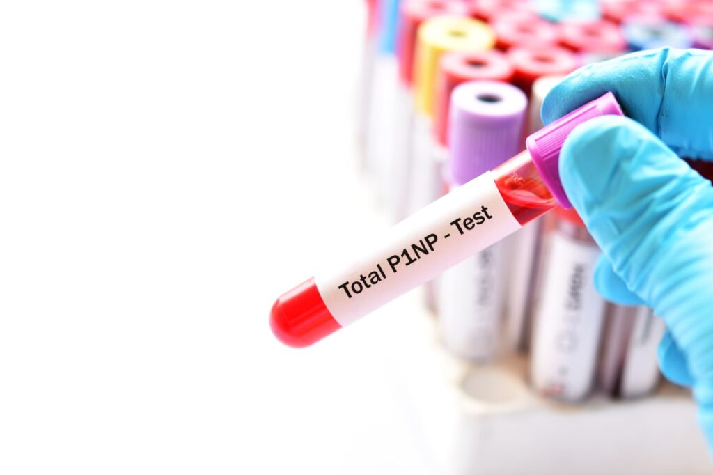
Bone Turnover Markers
Bone turnover markers are blood tests that measure the rate of bone resorption (osteoclast activity, measured by CTx) and bone formation (osteoblast activity, measured by P1NP) in real time. Unlike DEXA or REMS, which give you a snapshot, bone markers tell you about the direction your bone health is heading. Unlike your bone, which takes years to show change, the bone cells themselves show change within a few months.
This means you can tell whether your exercise program, nutrition plan, or medication is actually moving the dial, without waiting two years, or longer for a follow-up DEXA.
Key Takeaway
If you want more detail about your bone health, ask your DEXA provider about adding TBS and VFA to your next scan. Both are evidence-based, guideline-supported, and provide information your physician can act on immediately. Bone turnover markers through a simple blood test add yet another layer of insight.
Is REMS a Good Alternative to a DEXA Scan?
REMS is best understood as a supplement to DEXA rather than a replacement for it. It is a genuine alternative in specific situations: when a patient cannot be transported to a fixed DEXA unit, when radiation must be avoided, when spinal hardware or severe degenerative change makes DEXA readings unreliable, or when no DEXA is accessible at all. Outside those situations, DEXA remains the test that clinicians use to diagnose osteoporosis and decide on treatment, and REMS results will not usually change a treatment decision.
If you are looking for an alternative because you want to avoid radiation, it is worth knowing that a DEXA scan delivers roughly the same radiation dose as one day of ordinary background exposure. For most people that is not a reason to avoid it.
The Bottom Line
REMS is a creative technology with genuine appeal, especially for people who want a quick, radiation-free look at their bone health. It is not inherently bad, and you should not feel foolish if you’ve had one. But the latest research makes it clear that you should understand what it is and what it isn’t.
What it is:
A portable, radiation-free device that provides a demographic-weighted estimate of bone mineral density and fracture risk that correlates well with DEXA at a population level. It has specific, well-defined use cases, particularly when DEXA is physically impractical, when DEXA interpretation is compromised by hardware or degenerative changes, or when radiation must be avoided.
What it isn’t:
A direct, independent measurement of your individual skeleton that can reliably detect outliers, track treatment response, or replace DEXA for clinical decision-making.
If you’re serious about understanding your bone health, start with a DEXA. Ask about adding TBS and VFA. Consider bone turnover markers. And make sure you’re working with a clinician who can put all the pieces together into a plan that’s right for you.
Frequently Asked Questions About REMS Bone Scans
Is a REMS scan as accurate as a DEXA scan?
Population-level studies show REMS correlates well with DEXA. However, a 2026 study found that over 90% of the REMS-BMD result can be explained by the patient's age and weight alone — compared to 20–50% for DEXA. This means REMS may be less accurate for individuals whose bone health is significantly different from what their demographics would predict. DEXA remains the gold standard for osteoporosis diagnosis.
Is REMS (Echolight) FDA-approved?
REMS has received FDA 510(k) clearance, which means it has been cleared for marketing by demonstrating substantial equivalence to an existing device. This is a different regulatory pathway than "FDA approval," which involves a more rigorous premarket approval (PMA) process. Both DEXA devices and REMS hold 510(k) clearance.
How much does a REMS scan cost?
A REMS scan typically costs between $300 and $400 or more out of pocket. Insurance coverage is currently very limited. Some REMS providers have reported that Medicare may cover the scan in certain circumstances, but this is not guaranteed. In contrast, DEXA scans are widely covered by Medicare and most insurance plans.
Can I compare my REMS results to my DEXA results?
No. REMS and DEXA use fundamentally different technologies, and their T-scores and BMD values are not directly comparable. Differences between them do not mean one is "wrong." For tracking bone health over time, use the same technology — and ideally the same facility — for each scan.
Should I get a REMS scan instead of a DEXA?
In most cases, no. DEXA remains the clinical standard for osteoporosis diagnosis and treatment monitoring. REMS may be a reasonable option when DEXA is unavailable, unreliable (e.g., significant spinal arthritis or hardware), or when radiation must be avoided (e.g., pregnancy). It should not replace DEXA for treatment decisions.
Is the REMS Fragility Score a reliable measure of bone quality?
The Fragility Score is marketed as a measure of bone microarchitecture, but the Chan et al. 2026 study found that a simple mathematical function of age alone predicted over 95% of femoral neck Fragility Score variance and over 80% at the lumbar spine. This suggests it may reflect demographic weighting more than independent bone quality assessment.
Can REMS track bone density changes over time?
Early evidence is emerging. A 2026 study (Semeraro et al.) reported that REMS detected significant BMD increases at the hip after 6 months of Romosozumab (Evenity) therapy in 74 postmenopausal women, while an untreated control group showed stable readings. The authors confirmed that weight did not change during the study period, helping to address the concern that weight fluctuations could drive REMS changes. However, Romosozumab produces the largest and most rapid BMD gains of any osteoporosis drug, making it the easiest possible treatment effect to detect. Whether REMS can reliably detect the smaller, slower changes from more commonly prescribed medications like bisphosphonates or denosumab remains unproven. The study also lacked a parallel DEXA comparison. Until further evidence is available, be aware that a 5 kg weight change can shift REMS-BMD by approximately 4.3%, which may mask or mimic real bone changes.
Does Medicare cover a REMS scan?
Medicare coverage for REMS scans is not yet standard. Some providers report obtaining coverage in specific cases, but this varies by region and circumstance. DEXA bone density testing is a covered Medicare benefit with established guidelines.
Where can I get a REMS scan?
REMS/Echolight scanners are available at a growing number of wellness centers, chiropractic offices, and medical practices across the United States. The Echolight Medical website maintains a provider locator. Availability remains limited compared to DEXA, which is available at most hospitals, imaging centers, and many physician offices.
Does REMS give more accurate results for petite or small-framed women?
This is a common belief, but it is not supported by the evidence. DEXA can underestimate bone density in petite individuals because it measures areal density — a 2D measurement of a 3D structure — so smaller bones can appear less dense than they truly are. This is a well-known limitation. However, REMS does not solve this problem and may make it worse. Because REMS output is heavily influenced by the patient's weight, and petite women tend to weigh less, REMS may assign a lower bone density score based on the algorithm's demographic weighting rather than an actual measurement of thinner bone. If bone size is a genuine concern, ask your clinician about a BMAD (Bone Mineral Apparent Density) adjustment to your DEXA results, which partially corrects for vertebral size.
Which test better predicts real-world fracture risk — DEXA or REMS?
DEXA's ability to predict fractures has been validated in large studies spanning decades. The Study of Osteoporotic Fractures followed nearly 8,000 women and showed that a single DEXA measurement can predict hip fractures for up to 25 years. FRAX, the most widely used fracture risk calculator, was built on DEXA data. REMS has one published fracture prediction study (Pisani et al., 2023), conducted by researchers affiliated with Echolight, which found the Fragility Score predicted fractures over 5 years. However, the Chan et al. 2026 study found the Fragility Score is over 95% predicted by age alone — and since age is independently the strongest predictor of fracture, this correlation may reflect the well-established relationship between aging and fracture risk rather than an independent measurement of bone quality.
If BMD correlates with body weight, why aren't DEXA T-scores adjusted for weight?
DEXA T-scores compare your bone mineral density to a young adult reference population matched by sex and ethnicity, but not by weight or frame size. This is by design: the WHO threshold was calibrated to fracture risk, and lower body weight is independently associated with higher fracture risk. So while the T-score system may feel like it penalizes smaller people, it captures a real clinical signal. REMS uses a reference database stratified by BMI, which means it compares lighter individuals to other lighter individuals — producing more favourable T-scores. But this is also the demographic weighting that the Chan et al. study identified as problematic, because it may produce reassuring scores while masking genuine skeletal fragility caused by medication side effects, hormonal changes, or nutritional deficiencies. The safest approach is to discuss your T-score in context with your clinician and factor in your FRAX score.

Margaret Martin
Further Readings


References
- Chan D, Chen W, Yabsley E, Pocock N. Demographic determinants of REMS-derived BMD and fragility score. Osteoporos Int. 2026. doi:10.1007/s00198-026-07960-4
- Bobelyak M, Vaculik J, Stepan JJ. Bone mineral density assessment using radiofrequency echographic multispectrometry (REMS) in patients before and after total hip replacement. Osteoporos Int. 2025;36:2237–2244. doi:10.1007/s00198-025-07685-w
- Al-Daghri N, Brandi ML, Reginster JY, et al. Letter to the Editor [rebuttal]. Osteoporos Int. 2026;37:545–548. doi:10.1007/s00198-025-07817-2
- Bobelyak M, Vaculik J, Stepan J, Pocock N. Author response to OSIN-D-25-01882. Osteoporos Int. 2025. doi:10.1007/s00198-025-07789-3
- Zambito K, Kushchayeva Y, Bush A, et al. Proposed practice parameters for the performance of radiofrequency echographic multispectrometry (REMS) evaluations. Bone Jt Open. 2025;6:291–297. doi:10.1302/2633-1462.63.BJO-2024-0214.R1
- Fuggle NR, Reginster JY, Al-Daghri N, et al. Radiofrequency echographic multi spectrometry (REMS) in the diagnosis and management of osteoporosis: state of the art. Aging Clin Exp Res. 2024;36:135. doi:10.1007/s40520-024-02830-x
- Pisani P, Conversano F, Muratore M, et al. Fragility Score: a REMS-based indicator for the prediction of incident fragility fractures at 5 years. Aging Clin Exp Res. 2023;35:763–773.
- Di Paola M, Gatti D, Viapiana O, et al. Radiofrequency echographic multispectrometry compared with dual X-ray absorptiometry for osteoporosis diagnosis. Osteoporos Int. 2019;30:391–402.
- Blake GM, Fogelman I. Technical principles of dual energy X-ray absorptiometry. Semin Nucl Med. 1997;27:210–228.
- Semeraro A, Chialà A, Carafa A, Fanizzi R, Di Tano E, Palmisano M, Santoro C, Dibenedetto F, Napoli N. Very short-term monitoring of Romosozumab longitudinal effects in a cohort of postmenopausal women by means of Radiofrequency Echographic Multi-Spectrometry (REMS) technology. Aging Clin Exp Res. 2026;38:137. doi:10.1007/s40520-026-03391-7
Comments